
 USA
USA Australia
 Brazil
 China
China Croatia
 France
 Germany
 Italy
Italy Japan
Japan Latin America
 Netherlands
 Spain
Spain Turkey
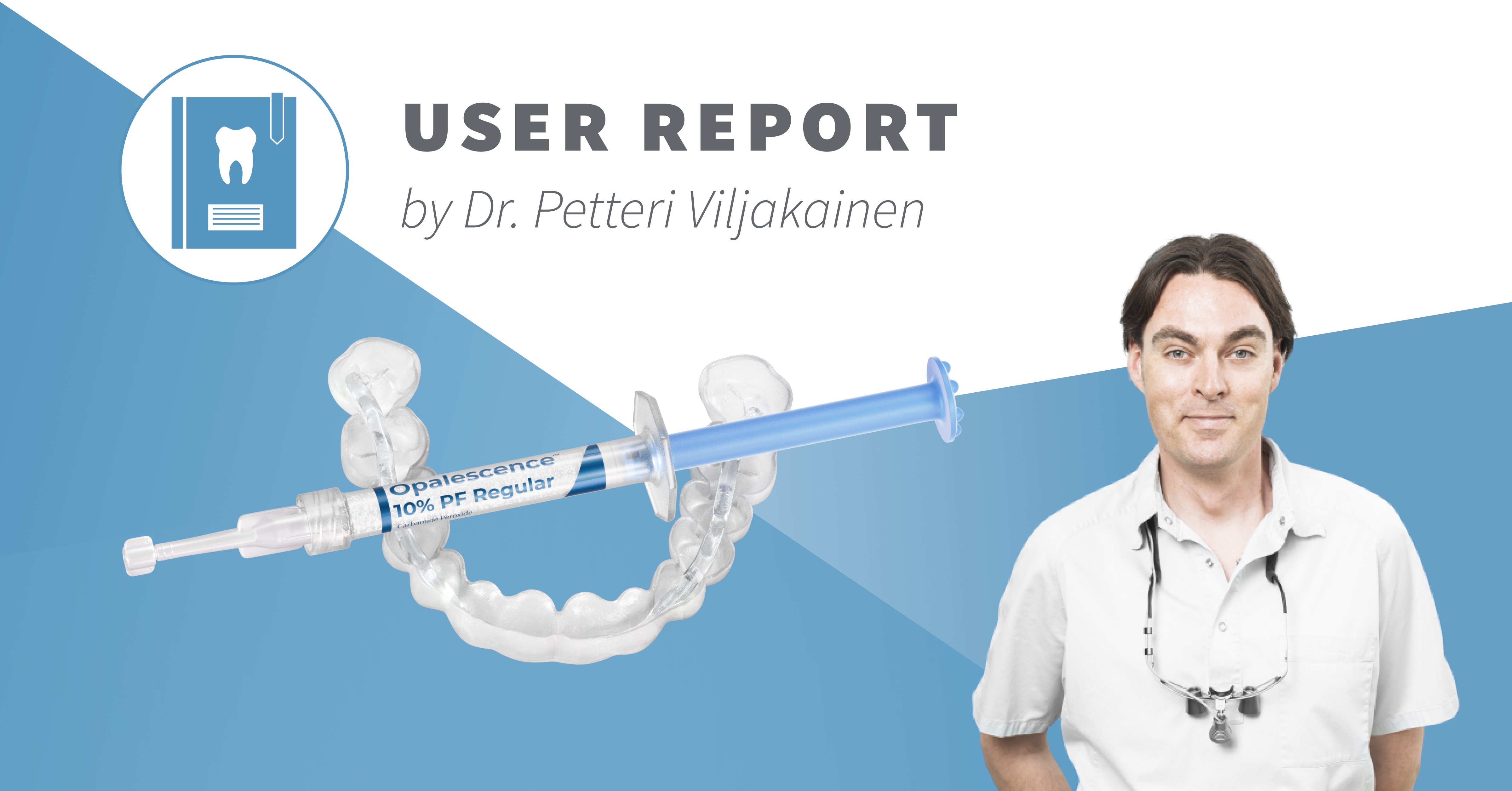


A user report by Dr. Petteri Viljakainen
Sometimes, discoloration of traumatized teeth can develop gradually and without noticeable symptoms, leading to diagnostic and therapeutic challenges at a later stage. The following case illustrates such a scenario of a 28-year-old female patient, who needed a treatment effectively nineteen years after the initial trauma.
She had suffered from a dental trauma in 2006, when she was only nine years old. According to her, the affected tooth 11 appeared completely normal for many years after the incident. Around 2016, however, she first noticed a slight color change, and by 2018 the discoloration had become increasingly significant (Fig. 1).

Fig. 1 – Initial situation
After having completed her studies in Dentistry, she assessed the situation herself in 2025. Radiographic examination revealed a completely obliterated pulp chamber, with no visible canal structure, although there were no radiographic signs of any periapical pathology or bone resorption (Fig. 2). Consequently, neither a root canal treatment nor an internal whitening were feasible. Additionally, the tooth did not react to vitality tests, but there were no clear indications of pulpal necrosis.

Fig. 2 – X-ray image
These findings led her to the assumption that a veneer would be the only solution, one which she would not like to pursue if any alternatives were available. She finally presented herself in my practice, seeking for a minimally invasive treatment.
Because of the already mentioned complete canal obliteration, and the impossibility of conventional endodontic therapy, Internal bleaching was equally excluded, because accessing the pulp chamber would have required an invasive intervention.
As there were no radiographic or clinical signs of inflammation (Fig. 2), I decided to follow an external whitening protocol to achieve aesthetic improvement without compromising the tooth structure.
Following a professional tooth cleaning, I took the tooth colour which turned out to be an A3 at the affected tooth 11, alongside a B1 at tooth 21 (Figs. 3 and 4).

Fig. 3 – Initial shade: tooth 11

Fig. 4 – Initial shade: tooth 21
For the whitening procedure we used an already existing custom-tray and Opalescence™ PF whitening gel with 10% carbamide peroxide (Ultradent Products, Inc.). First, the upper jaw was treated for two weeks to address the overall surrounding tooth color (Fig. 5).

Fig.5 – Result after 3 weeks: upper jaw
Subsequently, the treatment focused solely on the affected tooth 11 for an additional period of three weeks. After five weeks in total, the tooth shade had improved to a beautiful BL4 at the 11 and BL3 at 21 (Fig. 6 and 7), which means that the tooth colour of 11 almost matched the surrounding teeth (Fig. 8). The patient was fully satisfied with the outcome and delighted that no invasive intervention was necessary.

Fig. 6 – final result after 5 weeks: tooth 11

Fig. 7 – Final result after 5 weeks: tooth 21

Fig. 8 – Final result after 5 weeks: upper jaw
It was equally rewarding for me to observe that a classical external whitening procedure using a relatively low-concentration gel could achieve such a great whitening effect. Considering that the next alternative would have been possibly an indirect veneer, this treatment clearly turned out to be the better option. Even if the external whitening protocol would not have achieved the desired result, it would still be important to consider, as any shade improvement can only facilitate the work of the dental technician for the ceramic making.
This case specifically demonstrates that opting for more gentle, less invasive treatments can be worthwhile, even if the outcome might be uncertain initially. The excellent result achieved with Opalescence PF 10% highlights how effective and reliable external whitening protocols can be, even in complex cases with complete pulp canal obliteration. The gentle formulation provided a remarkable effect without compromising the tooth structure or causing sensitivity. Most impressively, even the patient herself – a young dentist – was delighted by the aesthetic result and the fact that this simple, conservative approach could deliver such a convincing outcome. Ultimately, the case shows that less can definitely be more in modern dentistry.
About the User:

Dentist Petteri Viljakainen
- Graduation, University Helsinki, Finland in 2009
- Dentist and partner at the eSmile practices in Espoo and Helsinki, Finland
- focused on cosmetic dentistry
- Focus & Special Interests:
- Cosmetic and aesthetic dentistry, with continuous participations in lectures and seminars worldwide
- Memberships:
- Accredited member of the American Academy of Cosmetic Dentistry (AACD)
- Accredited member of the British Academy of Cosmetic Dentistry (BACD)
- Accredited member of the IAS Academy
- Other activities:
- Key Opinion Leader for leading dental manufacturers
- International lecturer and hands-on trainer on dental materials and techniques as well as on dental photography